Member-only story
CD5-Negative, CCND1-IGH Translocation- Negative Mantle Cell Lymphoma
Lessons From the Friday Unknowns
Histologic sections show needle–shaped fragments of lymphoma.

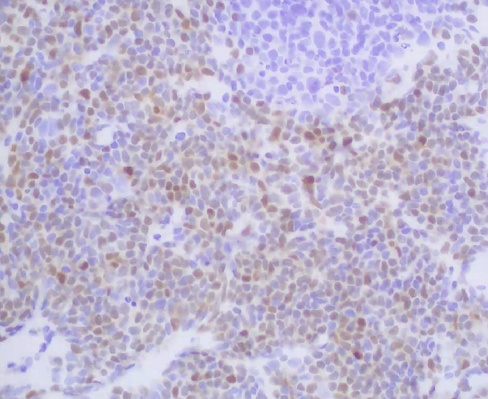
The neoplasm has a vaguely nodular and diffuse pattern and is composed predominantly of small lymphoid cells with slightly irregular nuclear contours.

There are many blood vessels with sclerotic walls. A few residual reactive germinal centers are present surrounded by the neoplasm.

In some areas, plasma cells are present. It is unclear if these cells are a part of the neoplastic process.

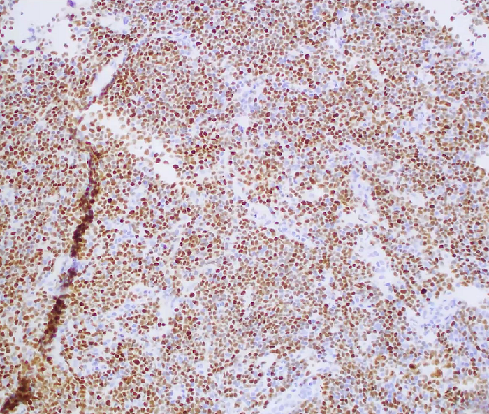
The neoplastic cells are positive for CD20, PAX–5, BCL–2, Mum 1/IRF4 (weak, subset), cyclin D1 (strong) and SOX-11 (strong) and are negative for CD3, CD5, CD10, CD23, BCL–6, TdT, C–MYC, CD138.
The antibody specific for Ki-67 shows a proliferation rate of approximately 10%.

Flow cytometry immunophenotypic studies show that the neoplastic cells are positive for monotypic surface lambda, CD19, CD20, CD43 (+/-), and CD45, and are negative for CD5, CD10, CD11c, CD23, CD25, CD71, CD103 and CD123.
This case was assessed by fluorescence in situ hybridization analysis to assess for CCND1–IGH translocation: The translocation was not detected.