Member-only story
CD5-Negative Mantle Cell Lymphoma, with Classic Histology & Increased Proliferation Index
Lessons From the Friday Unknowns
Histologic features of this lymphoma are unusual. Lymphoma cells are monomorphic appearing cells of small-intermediate size with irregular nuclear contours and condensed nuclear chromatin pattern. In routinely H&E stained sections, a nodular pattern is recognized in some areas.


Immunoperoxidase stained sections show positive immunoperoxidase staining of lymphoma cells for SOX11, Cyclin-D1, CD20, PAX-5 and Bcl-2. In focal areas and particularly in areas where a nodular pattern is suggested, a subset of lymphocytes shows dimly positive immunostaining for CD10. In multifocal areas and particularly in areas where a nodular pattern is present, a subset of lymphocytes shows positive immunostaining for BCL-6. Nodular meshworks of CD21-immunoreactive follicular dendritic cells highlight the nodular pattern pattern present in approximately one third of the lymphomatous tissue. Lymphoma cells are non-immunoreactive for CD3, CD5, c-MYC (5–10% of cells positive) and CD30.
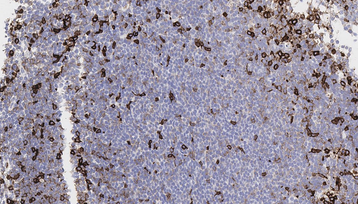
Immunoreactivity of lymphoma cells for Ki-67 antigen is variable, with highest immunoreactivity present in areas where a nodular pattern is suggested. The percentage of Ki-67-positive lymphoma cells ranges from 15–20% to as much as 60%, averaging overall approximately 50%.
In situ hybridization for EBV (EBER) is negative.


Flow cytometric analysis showed showed an 89% CD10-dim positive, kappa-light chain restricted B-cell population. The immunophenotypic profile was reported as positive for CD19, CD20, CD10 (dim), CD38 (dim to negative), CD52, IcCD22 and kappa and as negative for CD5 and lambda.
FISH analysis is positive for IGH/MYEOV/CCND1 rearrangement and a negative result for IGH/BCL2 rearrangement.
Link to digital slides: bit.ly/3Hi2bLG | Case 4