Member-only story
High Grade B-Cell Lymphoma with MYC & BCL2 Gene Rearrangements
Lesson From the Friday Unknowns
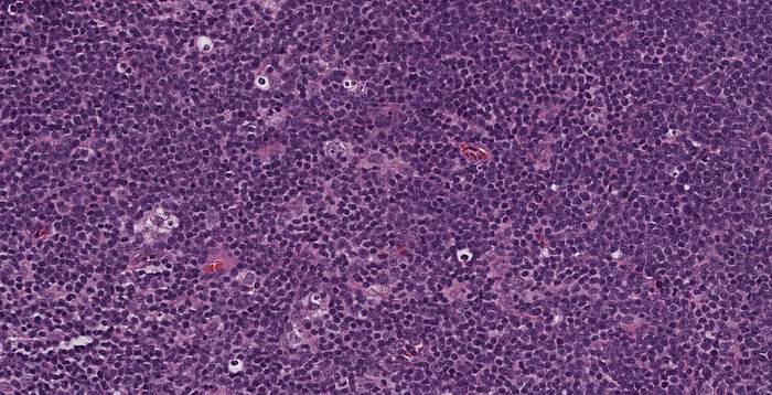
Lymphoma cells are of intermediate-large size. Mitotic division figures are present and high number within the lymphomatous proliferation. Approximately half of the lymphomatous proliferation has follicular pattern and at least half has diffuse pattern. In most areas of follicular lymphoma, the neoplastic follicles comprise sheets of centroblasts, while in a few areas of follicular lymphoma, the neoplastic follicles comprise a mixed population of centrocytes and centroblasts (grade 3A and grade 1–2).


Immunoperoxidase stained sections show positive immunoperoxidase staining of lymphoma cells for CD20, PAX-5, BCL6, Bcl-2, CD10 and MYC protein, with non-immunoreactivity of lymphoma cells for CD3, CD5, CD43, MUM-1, CD23, CD30 and cytokeratin AE1/AE3. Proliferation of lymphoma cells, as determined by immunohistologic staining for Ki-67, is high, with percentage of Ki-67 positive cells ranging from 70% to 100%. The follicular pattern is highlighted by the presence of nodular meshwork’s of CD21-positive follicular dendritic cells.
Fluorescence in situ hybridization for EBV (EBER) was negative.
Flow cytometric analysis detected abnormal B cells with mixed/increased cell size, positive for CD19, CD10, CD20, CD38, CD43, FMC7 and cytoplasmic kappa immunoglobulin light chain and negative for CD5, CD23, CD25, CD30 and surface immunoglobulin light chain.
By report of Integrated Oncology, FISH analysis gave a positive result for MYC gene rearrangement and BCL-2 gene rearrangement, while no rearrangement of BCL6 was detected.
Link to digital slides: bit.ly/3LxEvDc | Case 1
