Member-only story
Inflammatory Myofibroblastic Tumor
Lessons From the Friday Unknowns
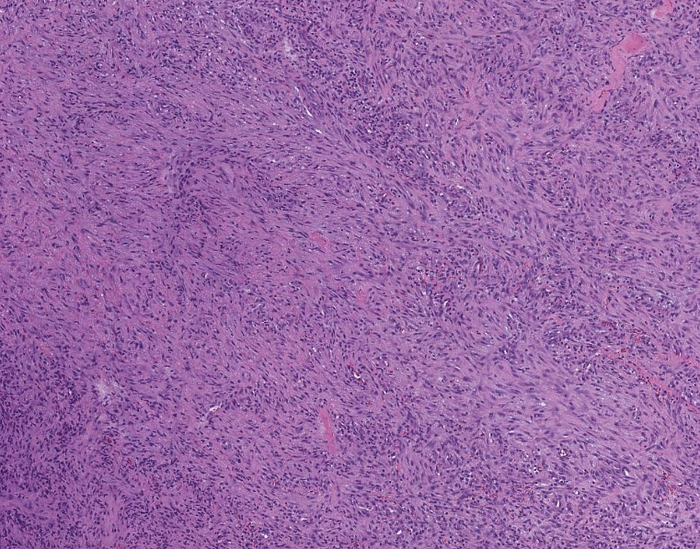
Histologic sections show a lesion with extensive coagulative necrosis. There is no evidence lymph node in this specimen.

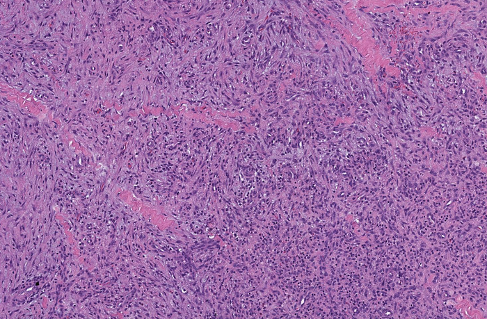
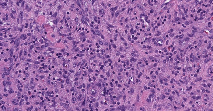
The lesion is composed of numerous spindle cells associated with inflammatory cells including lymphocytes, eosinophils and neutrophils, the latter near the areas of necrosis.


The spindle cells are arranged in loose fascicles or are more spread out and are arranged in a stellate fashion resembling, in part, granulation tissue. Mitotic figures are identified.

The spindle cells are positive for smooth muscle actin, CD10 and BCL-6 (moderate), and are negative for CD1a, CD3, CD5, CD20, CD21, CD30, CD31, CD34, CD68, PAX-5, BCL-2, cyclin D1, keratin (AE1/AE3), desmin, S100 protein, HHV8 and STAT6. The antibody specific for Ki-67 highlights…
