Member-only story
Marginal Zone Lymphoma with Residual Germinal Centers
3 min readAug 28, 2021
Lessons From the Friday Unknowns
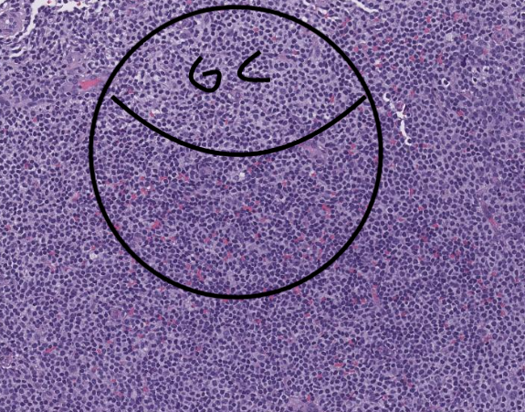
Histologic sections show adipose tissue extensively involved by lymphoma.

The neoplasm has a diffuse pattern and is composed of small cells with abundant pale cytoplasm suggestive of marginal zone differentiation. The lymphoma has very few mitotic figures and there are no areas of necrosis.


There are some areas of larger cells representing residual follicle centers surrounded by and partially infiltrated by the neoplasm.


