Member-only story
Nodular Lymphocyte Predominant Hodgkin Lymphoma
Lessons from the Friday Unknowns
Sections of the right cervical lymph node reveal the presence of vaguely nodular clusters of atypical large lymphoid cells having slightly lobulated nuclei with a vesicular chromatin pattern and distinct nucleoli and moderate pale staining cytoplasm, consistent with LP (L&H) cells.
The atypical large lymphoid cells are surrounded by lymphocytes and scattered histiocytes. A vaguely nodular growth pattern is evident in routinely stained sections.

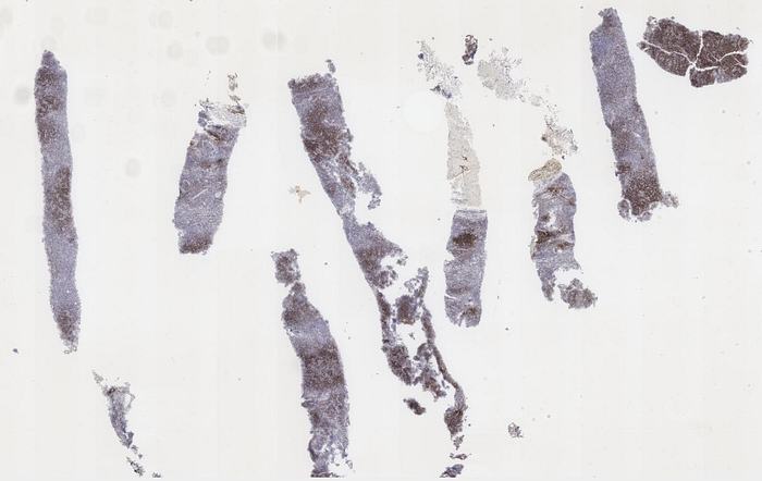
Submitted immunoperoxidase stained sections show positive immunoperoxidase staining of the atypical large lymphoid cells for CD20, with non-immunoreactivity of the cells for Bcl-2. The background lymphocytes are a mixed population of CD20 positive and CD3 positive cells. In the vaguely nodular areas where the atypical large, CD20-positive lymphoid cells are present, small CD20 positive lymphocytes are increased, while CD3-positive lymphocytes are relatively more numerous in the intervening areas of the tissue between the nodules. Within the lymphomatous nodules, CD3 positive lymphocytes are tightly clustered around the atypical large B cells and appear microscopically to be somewhat larger than T cells present in areas between the nodules, suggesting that they represent activated T cells. The immunoperoxidase stain for CD23 reveals the presence of loose, vaguely nodular meshwork’s of follicular dendritic cells within the lymphomatous nodules.


A history of nodular lymphocyte-predominant Hodgkin lymphoma, established on biopsy of a submandibular lymph node, is provided by clinic notes.
Link to digital slides: https://bit.ly/3DyDfxK | Case 2