Member-only story
Primary Cutaneous Marginal Zone Lymphoma
Neoplasm Lessons From the Friday Unknowns
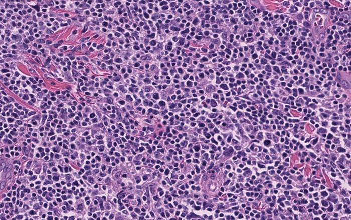
Histologic sections of the excision specimen show lymphoma involving the superficial and deep dermis and extending into the subcutaneous adipose tissue. The overlying epidermis is not involved. The infiltrate is composed of numerous small lymphoid cells with areas of increased large cells. Some of these large cells may represent reactive follicle centers surrounded and infiltrated by the infiltrate.


The neoplastic cells are positive for CD20 and are negative for CD3, CD4, CD5, CD8, CD10, CD21 and CD23. The antibody specific for CD30 highlights occasional large cells representing approximately 10% of all cells in the specimen. The antibody specific for cyclin D1 weakly highlights endothelial cells, epithelial cells, histiocytes and possible a small subset of large lymphoid cells.The antibody specific for Ki-67 shows a proliferation rate of approximately 15%.
